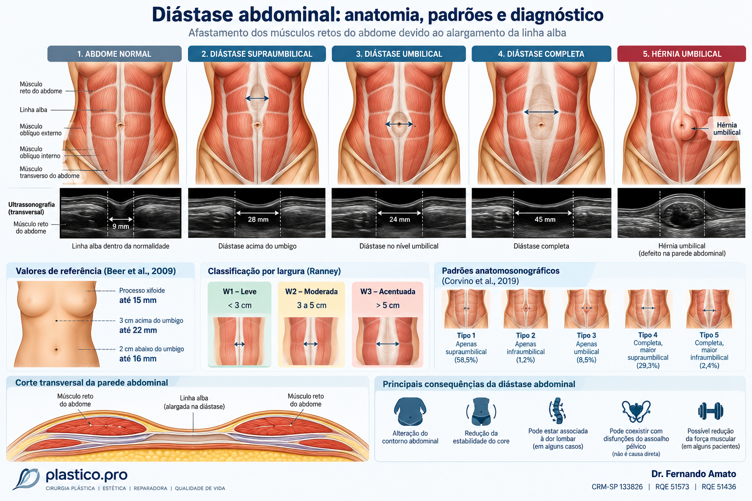
What is diastasis recti?
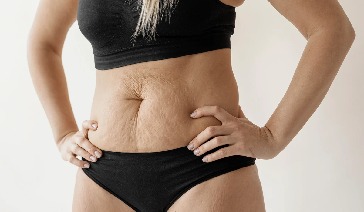
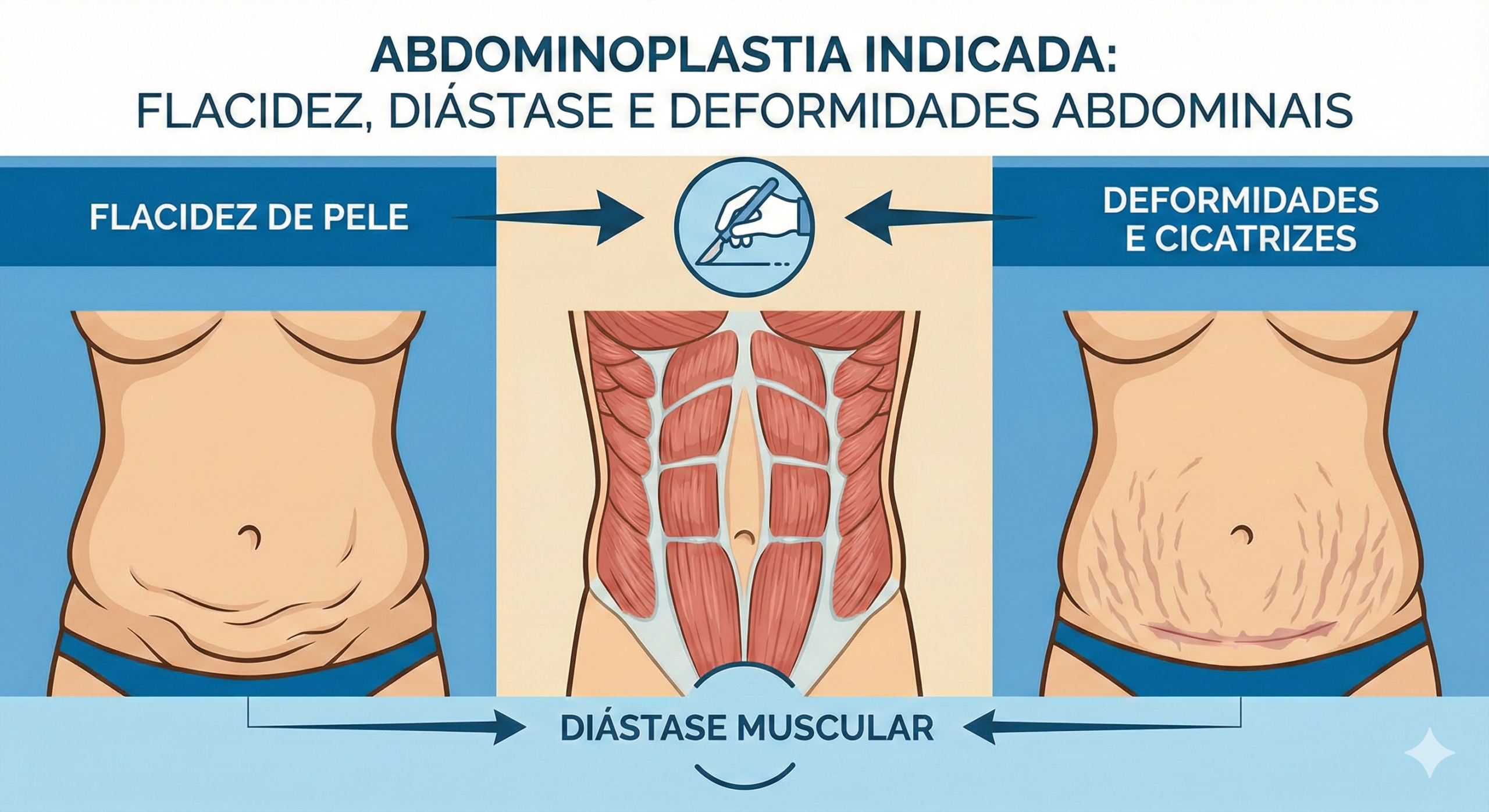
Diastasis recti (diastasis of the rectus abdominis muscles) is characterized by the separation of the muscles of the anterior abdominal wall due to the widening of the linea alba, the fibrous structure that connects them. It can produce a central bulge, especially during exertion. It is common after pregnancy, but it also occurs in men and in women without a history of pregnancy.
Although abdominal contour is a common complaint, it is important to evaluate possible symptoms and functional changes separately. Not all diastasis requires surgery.
What is the normal distance between the abdominal muscles?
There is a physiological separation between the rectus muscles. Beer and colleagues (2009) evaluated 150 nulliparous women using ultrasonography and found upper reference limits that varied according to anatomical level:
| Measurement site | Reference limit |
|---|---|
| Xiphoid process | 15 mm |
| 3 cm above the umbilicus | 22 mm |
| 2 cm below the umbilicus | 16 mm |
The European Hernia Society (EHS) proposes defining diastasis as an inter-rectus distance greater than 2 cm. This measurement alone does not represent an indication for surgical treatment: symptoms, location, function, and individual characteristics must also be evaluated.
Classification of diastasis by width
A traditional classification, attributed to Ranney, divides muscle separation into three grades:
- W1 (mild): less than 3 cm.
- W2 (moderate): between 3 and 5 cm.
- W3 (severe): greater than 5 cm.
Because the W1 category includes physiological distances, its interpretation must consider the diagnostic definition and the clinical context. The width measurement is not equivalent to functional severity.
Can diastasis have different distributions?
Yes. The linea alba can be widened above the umbilicus, only in the umbilical region, below the umbilicus, or along the entire abdomen.
Corvino and colleagues (2019) described five anatomical patterns in an ultrasound study. Among the 82 patients with diastasis in the sample, the distribution was:
| Pattern | Frequency in the sample |
|---|---|
| Supraumbilical only | 58.5% |
| Complete, with supraumbilical predominance | 29.3% |
| Umbilical only | 8.5% |
| Complete, with infraumbilical predominance | 2.4% |
| Infraumbilical only | 1.2% |
These percentages describe that specific sample and should not be generalized to the entire population.
Diagnosis by physical examination
Medical evaluation considers the abdominal contour, skin elasticity, previous scars, potential hernia, and midline protrusion during contraction.
The doctor may ask the patient, lying down, to lift their head and shoulders to highlight a longitudinal bulge. Palpation and clinical measurements help characterize the alteration, although they do not always allow for an accurate assessment of the full extent of the diastasis.
Ultrasound: when to use it?
Abdominal wall ultrasound is a useful complementary method because it does not use radiation, allows measurement of the distance between the rectus muscles at defined levels, and can evaluate the behavior of the wall during different maneuvers. It also helps detect associated hernias.
Mendes, Nahas, and colleagues (2007) compared ultrasound with intraoperative measurements during abdominoplasties. They found good correlation in the supraumbilical and umbilical regions; in the infraumbilical segment, ultrasound measurements were smaller than surgical ones.
To monitor results, it is important to standardize the site, position, and muscle condition during measurement. The interpretation of the exam must be integrated with the physical examination.
When to order a CT scan or MRI?
Computed tomography can help when there is doubt about hernias, complex abdominal wall alterations, or the need for reconstructive planning. Magnetic resonance imaging provides details on soft tissues and can be used in specific situations.
These exams are not routinely necessary for all patients with diastasis. Clinical evaluation and ultrasound generally suffice for initial investigation.
Is diastasis the same thing as a hernia?
No. Diastasis is the widening of the linea alba with separation of the rectus muscles, without requiring a fascial defect in the abdominal wall. A hernia implies a defect through which tissues can protrude.
The conditions can coexist. The presence of a hernia is relevant because it changes the surgical plan and may require specific techniques, with the potential use of mesh. It is not appropriate to present a universal percentage of hernias in patients with diastasis without defining the study population.
Can diastasis reduce abdominal strength?
The rectus, oblique, and transversus muscles participate in trunk movement and stabilization. The linea alba transmits tensions between them, and its widening can alter this distribution.
Some patients report weakness, difficulty with exercise, or functional discomfort. However, the width of the diastasis alone does not predict the severity of these symptoms.
Studies use dynamometry to measure strength and torque—the capacity to produce rotational force. Emanuelsson and colleagues (2016) evaluated surgical treatments and an exercise training group, finding functional improvements in selected patients. These results do not mean that surgery is superior to physical therapy for everyone.
Does diastasis cause low back pain?
The association is not consistently proven. A 2023 systematic review gathered 13 studies involving 2,820 participants: eight observed no significant association between diastasis and low back pain, and five found an association.
It is plausible that abdominal wall mechanics contribute to symptoms in certain patients, but one should not claim that every diastasis causes low back pain, nor promise that correcting it will eliminate the pain.
Is there a link to urinary incontinence?
Abdominal diastasis and pelvic floor dysfunctions can coexist, especially after pregnancy. However, association does not imply causation. Age, pregnancy, childbirth, and other factors also influence urinary and pelvic symptoms. Each condition must be investigated individually.
Does collagen play a role in the development of diastasis?
The linea alba contains collagen, an important component for the strength and organization of connective tissue.
Blotta and colleagues (2018) compared linea alba samples from 18 women with diastasis and 18 without the condition, finding lower amounts of type I and type III collagen in the group with diastasis. This finding suggests involvement of the extracellular matrix, but this small observational study does not establish a single cause.
Can physical therapy help?
Physical therapy may include abdominal strengthening, core stability training, breathing strategies, and pelvic floor rehabilitation when indicated.
Recent reviews show that some postpartum exercise programs can reduce the inter-recti distance, although the certainty of the evidence is limited. Specific protocols yield different results. Functional improvement can also occur without complete anatomical closure of the separation.
When should surgery be considered?
The decision depends on an individualized evaluation. Persistent functional symptoms, impact on quality of life, insufficient response to conservative treatment, presence of a hernia, excess skin, and overall clinical condition may all be relevant factors.
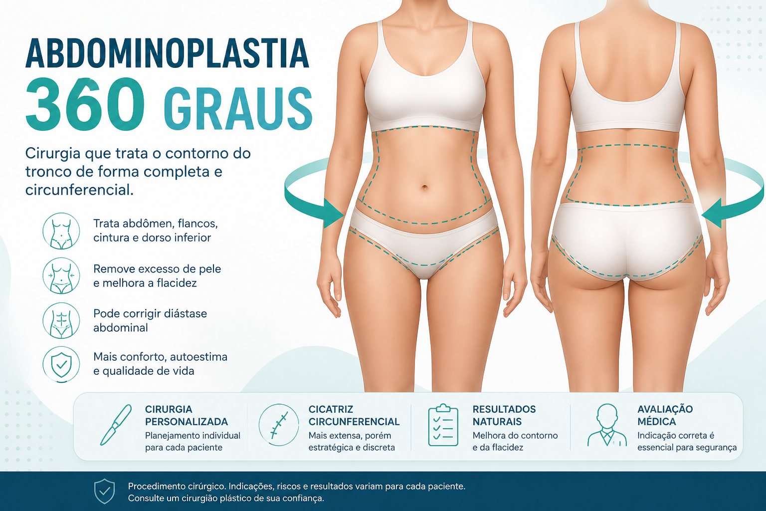
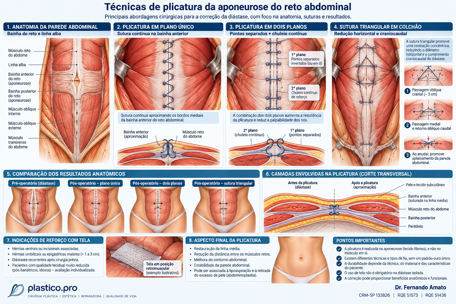
Surgical repair may involve plication, a suturing technique that brings together and reinforces the aponeurotic structure. When significant skin laxity is present, it can be combined with an abdominoplasty. Concomitant hernias require specific assessment.
As with any surgery, there are risks of seroma, hematoma, infection, healing abnormalities, recurrence, and thromboembolism. Universal functional improvement should not be promised.
Does surgery improve quality of life?
Olsson and colleagues (2019) followed 60 women with persistent symptomatic diastasis despite physical training. After surgical correction, they observed improvements in function and quality of life. The observational design does not allow for the conclusion of universal superiority of surgery compared to continuing conservative treatment.
Conclusion
Abdominal diastasis is an anatomical alteration of the linea alba that can affect body contour and, in some patients, be associated with functional symptoms. Diagnosis integrates clinical history, physical examination, and, when necessary, ultrasonography. The measurement of the inter-recti distance alone does not determine the need for surgery. Treatment must consider symptoms, anatomy, risks, expectations, and conservative alternatives.
Scientific references
- Hernández-Granados P, et al. European Hernia Society guidelines on management of rectus diastasis. Br J Surg. 2021;108(10):1189-1191. https://doi.org/10.1093/bjs/znab128
- Beer GM, et al. The normal width of the linea alba in nulliparous women. Clin Anat. 2009;22(6):706-711. https://doi.org/10.1002/ca.20836
- Reinpold W, et al. Classification of Rectus Diastasis—A Proposal by the German Hernia Society and the International Endohernia Society. Front Surg. 2019;6:1. https://doi.org/10.3389/fsurg.2019.00001
- Corvino A, et al. Diastasis of rectus abdominis muscles: patterns of anatomical variation as demonstrated by ultrasound. Pol J Radiol. 2019;84:e542-e548. https://doi.org/10.5114/pjr.2019.91303
- Mendes DA, et al. Ultrasonography for measuring rectus abdominis muscles diastasis. Acta Cir Bras. 2007;22(3):182-186. https://doi.org/10.1590/S0102-86502007000300005
- Emanuelsson P, et al. Operative correction of abdominal rectus diastasis reduces pain and improves abdominal wall muscle strength. Surgery. 2016;160(5):1367-1375. https://doi.org/10.1016/j.surg.2016.05.035
- Sokunbi G, et al. Is Diastasis Recti Abdominis Associated With Low Back Pain? A Systematic Review. World Neurosurg. 2023;174:119-125. https://doi.org/10.1016/j.wneu.2023.03.014
- Blotta RM, et al. Collagen I and III in women with diastasis recti. Clinics. 2018;73:e319. https://doi.org/10.6061/clinics/2018/e319
- Olsson A, et al. Cohort study of the effect of surgical repair of symptomatic diastasis recti abdominis on abdominal trunk function and quality of life. BJS Open. 2019;3(6):750-758. https://doi.org/10.1002/bjs5.50213
- Beamish NF, et al. Impact of postpartum exercise on pelvic floor disorders and diastasis recti abdominis: a systematic review and meta-analysis. Br J Sports Med. 2025;59:562-575. https://doi.org/10.1136/bjsports-2024-108619
- Lyons G, et al. What is the evidence for abdominal and pelvic floor muscle training to treat diastasis recti abdominis postpartum? Hernia. 2026. https://doi.org/10.1007/s10029-026-03660-4
Dr. Fernando Amato
Plastic Surgeon — CRM-SP 133826 | RQE 51573 | RQE 51436
https://plastico.pro
Illustrative image generated by artificial intelligence — insert manually before publication.
Agendar consulta
Precisa de orientação personalizada?
Marque uma avaliação com o Dr. Fernando Amato e tire as suas dúvidas sobre cirurgia plástica.
Falar no WhatsApp